What Flavor/Subtype of Concussion Does Your Patient Have? Psychological Type Deep-dive
Which of the Concussion Subtypes are Driving Your Patient’s Symptoms? A Psychological Type Deep-dive Welcome to the last and final part of the deep dive into the 4 different mild…
What Flavor/Subtype of Concussion Does Your Patient Have? Visual/Vestibular (Vestibuloocular) Type Deep-Dive
Introduction Post Concussion Syndrome (PCS) is a complex disorder in which various common concussion symptoms — such as headaches, dizziness, and cognitive dysfunction — persist for weeks, months, or even…
What Flavor/Subtype of Concussion Does Your Patient Have? Autonomic Type Deep-Dive
Concussions are often called invisible injuries, leaving patients with a myriad of physical, cognitive, and emotional symptoms. One unifying driver between all these symptoms can be a deceptively simple problem:…
The majority of individuals, approximately 60-70%, who sustain a concussion will recover within 4 weeks. While many strive to be among this majority, about 30-40% of concussion patients will experience post-concussive symptoms (PCS) that persist beyond the typical metabolic healing period. (1,2,3,4,5)
Identifying the source of a patient’s symptoms, known as “symptom generators,” can be challenging due to the wide variability of symptoms following a concussion.
Without a clear plan for assessing PCS, clinicians may misinterpret patient symptoms for the wrong concussion subtype. Determining the precise “symptom generator” is crucial for accurate diagnosis and effective treatment.
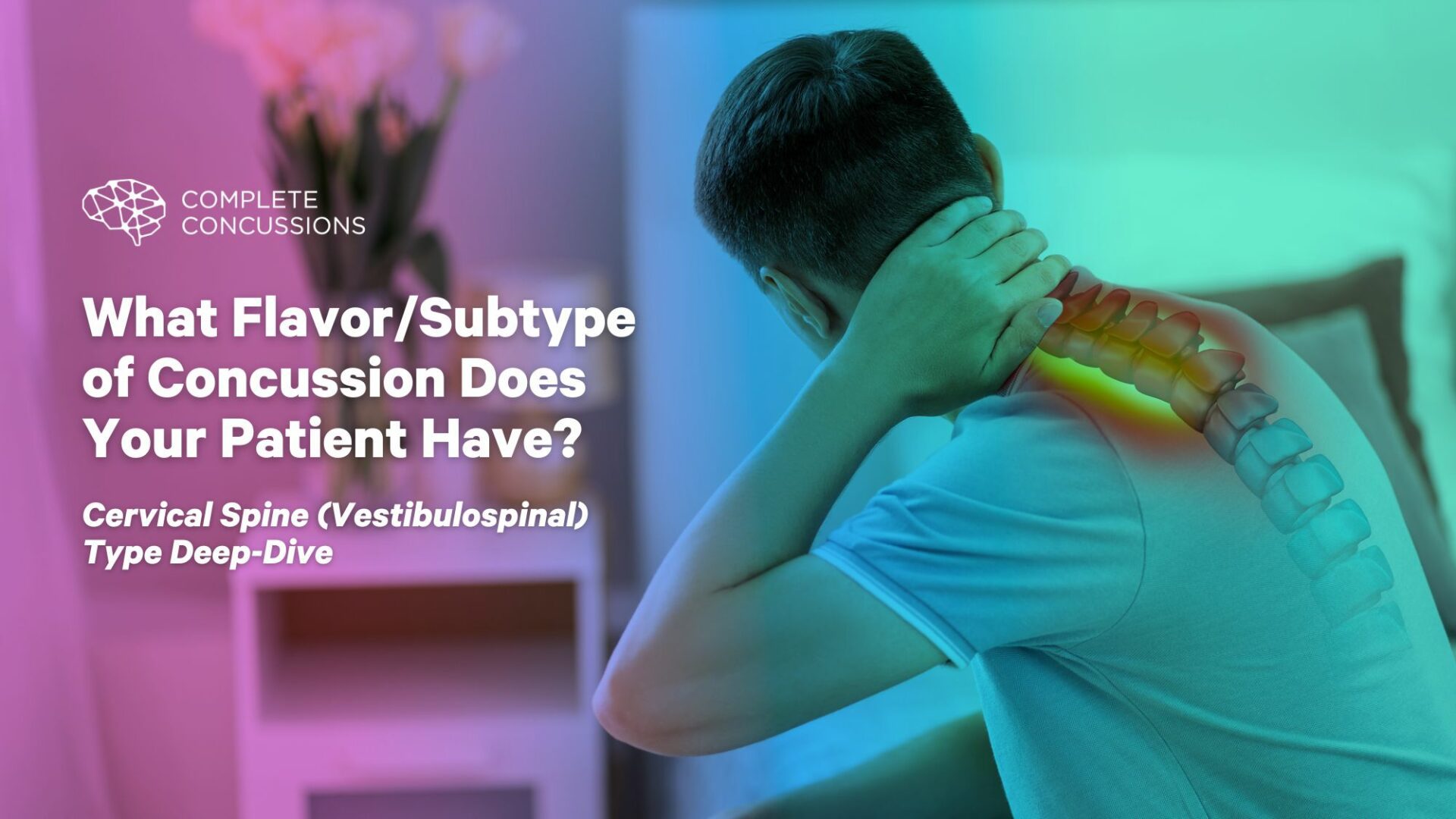
In this part of the series on concussion flavors, we will discuss the specific subtype related to the cervical spine. Our goal is to help you identify if your patient’s specific concussion subtype is one of cervical spine origin and develop a targeted treatment plan based on your findings.
Cervical Spine Involvement “The Cervicogenic Patient”.
Patients with a vestibulospinal or cervicogenic concussion “symptom generator” typically present with characteristic symptoms. Those with cervical spine involvement commonly report:
Imbalance
Lightheadedness
Cervical/Neck pain
Headaches that tend to be tension-type (rather than migrainous)
While these symptoms are common in patients with cervical dysfunction, they may also appear with other symptom generators, such as visual or autonomic dysfunction. Differentiating between these reported symptoms can provide the first clue in identifying the correct concussion subtype.
Patients may also provide insights through key triggers that provoke their symptoms. Those with a cervical spine subtype often report the following triggers:
Reported Triggers:
Head or neck movement
Worsening symptoms during exercise
Increased symptoms while driving
Why Does Cervical Dysfunction Occur Following Concussion?
#1 Cervical Sprain/Strain: Musculoskeletal Origin
The forces generated in a concussion injury (70-120 G’s) are sufficient to cause a cervical spine sprain or strain (4.5 G’s). (6)
Whiplash injuries and concussions are closely related in terms of symptom presentation. In fact, if clinicians were unaware of the specific injury, it would be nearly impossible to differentiate between the two based on symptoms alone.
This similarity arises from dysfunction in the cervical spine, particularly in the suboccipital region and surrounding neck muscles. This leads to a decreased range of motion, increased muscular tension, and proprioceptive dysfunction.
Common Concussion AND Whiplash Symptoms
Neck pain
Neck tightness/ decreased range of motion
Dizziness
Blurred vision
Mental/Cognitive fatigue
Headache
Memory/Concentration Impairment
#2 Cervical Proprioceptive Neural Injury
The upper cervical spine houses millions of proprioceptive organs densely populated within the facet joints and surrounding musculature. These proprioceptors relay constant information to the brain about the head’s position in space.
The intricate connection between neck muscles, joint proprioception, and other mechanisms means that injury to these structures can result in proprioceptive misinformation sent to the brain, leading to disequilibrium, dizziness, and balance issues. (7)
#3 Structural Integrity
With head injuries, the cervical spine can be impacted to an extent that poses a significant risk to its structural integrity. This includes the potential for cervical fractures, ligamentous injuries, and vascular incidents, all of which should be carefully considered.
Following head and cervical spine trauma, there’s a possibility of cervical spine instability and other injuries, such as occipital neuralgia, emerging. Therefore, special attention should be given to assessing and addressing these potential complications.
Cervical Spine Assessment.
When assessing a patient suspected of having cervical spine involvement, adopting a systematic approach is crucial. Before initiating a cervical spine assessment, it’s essential to consider the various dysfunctions that may be at play.
Conducting a comprehensive patient history can aid clinicians in identifying the specific mechanism of cervical spine dysfunction.
The three main categories outlined in the preceding section, “Why Does Cervical Dysfunction Occur,” encompass the typical presentations of patients with complaints related to the cervical spine.
Compromise to Structural Integrity (Ruling out red flags)
Musculoskeletal Injury
Proprioceptive Dysfunction
Structural Integrity and Red Flags
The close relationship between reported symptom cross-over, location, and implications for severe injury to the cervical spine should be thoroughly assessed before further evaluation and treatment can start.
Some key structures require specific attention when assessing the cervical spine for structural integrity and red flags.
Fracture (Canadian C Spine Rules)
In assessing a patient for potential cervical spine fracture, clinicians should adhere to the “Canadian C Spine Rules.” These rules provide specific criteria for identifying suspected fracture risk in individuals following trauma. With a sensitivity of 99.4% and a specificity of 45.1%, the Canadian C Spine Rules are highly effective at ruling out fractures. If a fracture is suspected based on these criteria, the next step would be to refer the patient for radiographic imaging. (8)
Ligamentous Integrity
Spinal stability is a critical factor to consider following trauma, particularly when deciding whether to implement treatments such as manual therapy and rehabilitative movement.
Various tests are available to evaluate the structural integrity of the cervical spine. (9,10,11,12)
Right and Left Alar ligament test
Sharp Purser Test (Integrity of Transverse ligament)
Spurling’s Test (Cervical radiculopathy)
Vascular Incident and Cranial Nerves
Vascular incidents, such as post-traumatic fistulas or dissections, may arise following significant head or neck trauma. During the initial examination, auscultation for bruits over the temporal arteries, mastoids, carotids, and with closed eyes can help assess for vascular turbulence that may indicate such occurrences.
Irrespective of the concussion subtype, a thorough neurological assessment is essential for clinicians to diagnose or exclude the possibility of additional pathology. Trauma to the neck is a known cause of occipital neuralgia, and a detailed evaluation of the cranial nerves can reveal structural abnormalities that warrant further investigation. (12)
Musculoskeletal Injury
Symptoms of an isolated cervical spine injury and concussion exhibit a significant overlap, often presenting similarly. Musculoskeletal injuries can even induce concussion-related symptoms that many patients may not attribute to a neck injury, such as dizziness, a sense of being off-balance, mental fatigue, and visual dysfunction. (7)
Headaches and neck pain are the most common symptoms associated with a genuine cervical spine injury, often leading to the onset of other related symptoms.
During a physical examination of the cervical spine, emphasis should be placed on assessing range of motion and palpation for tenderness and muscle spasms in the neck. Suboccipital muscles frequently bear the brunt of trauma to the head and neck and warrant thorough examination.
Musculoskeletal Assessment
Cervical Range of Motion (Passive and Active)
Palpation of muscle tenderness and spasm
Specific trigger point palpation for pain referral patterns
Special Consideration For Headache Symptoms
Headaches are a common symptom to present after a concussion. Due to the high incidence of headaches, clinicians need to use clear physical examination techniques to differentiate headache subtypes. In 2016, Delphi et al. outlined the most effective techniques physical therapists employ to detect headaches. (13)
Delphi determined the following as the most effective methods for the detection of headaches
Craniocervical flexion test
Cervical flexion rotation test
Active range of cervical motion
Trigger point palpation
Muscle test of the shoulder girdle
Passive physiological intervertebral movements
Thoracic spine screening
Combined movement test
Cervicogenic Headache Test Cluster
Combining the above tests can effectively rule out specific cervical spine dysfunctions. In the context of diagnosing cervicogenic headaches, a cluster of tests has demonstrated strong accuracy and high reliability. (14-15)
Cervical flexion rotation test
Smooth pursuit neck torsion test (SPNTT)
Cervical-joint reposition error test (JPET)
Proprioceptive Injury
Given the dense concentration of proprioceptive structures in the cervical spine, particular attention should be paid if a patient exhibits dizziness, balance dysfunction, and disequilibrium sensations.(7)
The cervical-joint reposition error test (JPET) pinpoints dysfunction of the muscle spindles in the upper cervical spine.
Furthermore, the smooth-pursuit neck torsion test (SPNTT) is valuable for assessing cervico-ocular dysfunction.
Enhanced understanding of the mechanisms underlying a patient’s concussion subtype can greatly aid clinicians in developing a more precise and personalized treatment plan.
Cervical Spine Treatment.
The primary objective in treating patients with a concussion subtype involving cervical spine dysfunction is to restore the patient to a state of normal function in both cervical spine biomechanics and proprioceptive function.
In instances where cervical spine dysfunction involves a significant musculoskeletal component, manual therapy, and rehabilitation techniques can yield beneficial outcomes.
Chiropractors and physical therapists, with their specialized expertise and training, are well-equipped to facilitate recovery through manual therapy techniques such as:
Soft tissue/ Myofascial work
Joint Manipulation
Dry Needling
Trigger point therapy
Stabilization exercises
If a proprioceptive dysfunction is identified as significantly impacting the examination, clinicians can utilize various rehabilitative techniques to aid patients in reintegrating this system and fostering a more comprehensive recovery.
Treatment Options for Cervical Spine Proprioceptive Input
Cervical JPET (as rehab)
Smooth Pursuits
Smooth Pursuits with neck torsion (SPNTT as rehab)
Bess H Exercise
Walking with Head Turns exercise
Bottom Line.
Neck pain and headaches frequently accompany concussion injuries, often signifying cervical spine involvement. However, the cervical spine can elicit other less typical symptoms, confusing clinicians attempting to identify the true “symptom generator” in their patients’ cases.
Having a clear and accurate plan for evaluation and detection can significantly impact treatment times and patient outcomes.
REFERENCES
Zemek R, Barrowman N, Freedman SB, et al. Clinical Risk Score for Persistent Postconcussion Symptoms Among Children With Acute Concussion in the ED [published correction appears in JAMA. 2016 Jun 21;315(23):2624].
Grubenhoff JA, Currie D, Comstock RD, Juarez-Colunga E, Bajaj L, Kirkwood MW. Psychological Factors Associated with Delayed Symptom Resolution in Children with Concussion. J Pediatr. 2016;174:27-32.e1. doi:10.1016/j.jpeds.2016.03.027
Howell DR, Zemek R, Brilliant AN, Mannix RC, Master CL, Meehan WP 3rd. Identifying Persistent Postconcussion Symptom Risk in a Pediatric Sports Medicine Clinic. Am J Sports Med. 2018;46(13):3254-3261.
Ewing-Cobbs L, Cox CS Jr, Clark AE, Holubkov R, Keenan HT. Persistent Postconcussion Symptoms After Injury. Pediatrics. 2018;142(5):e20180939.
Kara, S., Crosswell, H., Forch, K., Cavadino, A., McGeown, J., & Fulcher, M. (2020). Less than half of patients recover within 2 weeks of injury after a sports-related mild traumatic brain injury: a 2-year prospective study. Clinical journal of sport medicine, 30(2), 96-101.
Slade, S. (2009). Feel the G’s: The Science of Gravity and G-forces. Capstone.
Jaumard NV, Welch WC, Winkelstein BA. Spinal facet joint biomechanics and mechanotransduction in normal, injury and degenerative conditions. J Biomech Eng. 2011;133(7):071010. doi:10.1115/1.4004493
Stiell IG, Wells GA, Vandemheen KL, et al. The Canadian C-spine rule for radiography in alert and stable trauma patients. JAMA. 2001;286(15):1841-1848. doi:10.1001/jama.286.15.1841
Osmotherly PG, Rivett DA, Rowe LJ. Construct validity of clinical tests for alar ligament integrity: an evaluation using magnetic resonance imaging. Phys Ther. 2012;92(5):718-725. doi:10.2522/ptj.20110261
Hutting N, Scholten-Peeters GG, Vijverman V, Keesenberg MD, Verhagen AP. Diagnostic accuracy of upper cervical spine instability tests: a systematic review. Phys Ther. 2013;93(12):1686-1695. doi:10.2522/ptj.20130186
Uitvlugt G, Indenbaum S. Clinical assessment of atlantoaxial instability using the Sharp-Purser test. Arthritis Rheum. 1988;31(7):918-922. doi:10.1002/art.1780310715
Choi I, Jeon SR. Neuralgias of the Head: Occipital Neuralgia. J Korean Med Sci. 2016;31(4):479-488. doi:10.3346/jkms.2016.31.4.479
Luedtke K, Boissonnault W, Caspersen N, et al. International consensus on the most useful physical examination tests used by physiotherapists for patients with headache: A Delphi study. Man Ther. 2016;23:17-24. doi:10.1016/j.math.2016.02.010
Hall TM, Robinson KW, Fujinawa O, Akasaka K, Pyne EA. Intertester reliability and diagnostic validity of the cervical flexion-rotation test. J Manipulative Physiol Ther. 2008;31(4):293-300. doi:10.1016/j.jmpt.2008.03.012
Rubio-Ochoa J, Benítez-Martínez J, Lluch E, Santacruz-Zaragozá S, Gómez-Contreras P, Cook CE. Physical examination tests for screening and diagnosis of cervicogenic headache: A systematic review. Man Ther. 2016;21:35-40. doi:10.1016/j.math.2015.09.008
Dr. Denner is a highly skilled Chiropractic Physician and licensed Athletic Trainer based in Charlotte, North Carolina. Specializing in sports medicine and rehabilitation, he utilizes his unique background in manual therapy, functional rehabilitation, joint manipulation, and neuromuscular stabilization to effectively treat a wide range of musculoskeletal conditions.
As the owner and treating physician at Denner Chiropractic & Performance, Dr. Denner brings extensive experience and a comprehensive approach to patient care. With a focus on personalized, functional treatment plans, he empowers his patients to take an active role in their recovery.