Concussions can happen to athletes of any age participating in all kinds of sports. From hard hits in hockey and football to accidents in gymnastics and soccer, concussions are an…
Greater Toronto Area’s Leading Multidisciplinary Clinics for Concussion Recovery
Concussions are a common form of traumatic brain injury, often caused by blows to the head. They can result in an array of physical, cognitive, and emotional symptoms that may…
Why You Should Never Skip the Neck in Concussion Evaluation & Rehab.
Every patient with a concussion is a patient with a neck injury. Let me explain. A concussion results when the brain undergoes such rapid acceleration-deceleration that shearing and stretching of…
Rethinking Concussion Recovery: The Case for Active Rehabilitation Concussions are more than just a bump on the head, or “getting your bell rung”; they are a type of mild traumatic…
The visual system is one of the most complex of the human body’s systems. It begins with two simple organs—the eyes—but its workings are far more intricate than just seeing. Much of the visual system’s processes are carried out automatically and in real-time, allowing us to take in information about the world around us without having to consciously think about it.
The visual system consists of the eyes, the muscles that control eye movement, cranial nerves that carry signals from the brain to the eyes (and vice versa), and different areas of the brain where information is processed. Proper functioning of this complex network allows us to interact with the world around us. Visual functions include detecting shapes and movement, perceiving color and depth, focusing on objects in our environment, and more.
The occipital lobe of the brain houses the primary visual cortex and is responsible for our conscious and detailed vision. In other words, identifying what we see before us. The parietal lobe processes visual information about spatial consciousness that helps orient where we are in the surrounding environment. Our frontal lobe is where the initiation of most voluntary eye movement takes place, such as when we track an object through space, or intentionally shift our eyes from one point to another. The temporal lobe processes auditory information about space and detail, and matches this with spatial visual information for such functions as object naming. The cerebellum dictates the accuracy of our eye movements and the steadiness of our visual fixation, and the midbrain integrates visual information with that of many other pathways such as the vestibular system and sensorimotor feedback from proprioceptors and muscle spindles. With concussion and brain injuries having the potential to affect any one or combination of these areas, it should not be surprising that many different elements of visual function can be impacted.
A Team Effort: Vision’s Connection to Other Pathways
The visual system does not operate in isolation, but rather works very closely with the vestibular system and somatosensory systems of the body to help gather information about the world around us, and where we fit within it. This information is then relayed to the superior colliculus of the midbrain along with information from our auditory system and autonomic nervous system, where these inputs are reconciled against one another. If one system is not functioning correctly, it may cause dysfunction in another. For example, if the visual system is providing the brain with different information than the vestibular system about the head’s position in space, patients may feel dizzy or disoriented due to this mismatched input.
Concussions and Vision
A concussion, also referred to as a mild traumatic brain injury, can occur in many different ways, such as a car accident, during contact sports, workplace injuries, or as the result of physical abuse. Visual problems are one of the most common symptoms of concussion, with as much as 70% of patients experiencing some alteration in visual function post-injury, according to some studies [1]. Visual symptoms following head injury and concussion can vary widely, and may include such complaints as headaches, light sensitivity, eye strain, blurred vision, trouble focusing, double vision, difficulty with eye tracking, and sensitivity to motion in the visual field, among others. Persistent visual symptoms are one of the most common contributors to post-concussion syndrome. As such, it is important for clinicians to understand the different types of vision tests that can be performed on patients with post concussion symptoms as part of a comprehensive physical exam, and know when to refer for more specialized visual assessments to ensure your patient gets the right treatment to support their recovery.
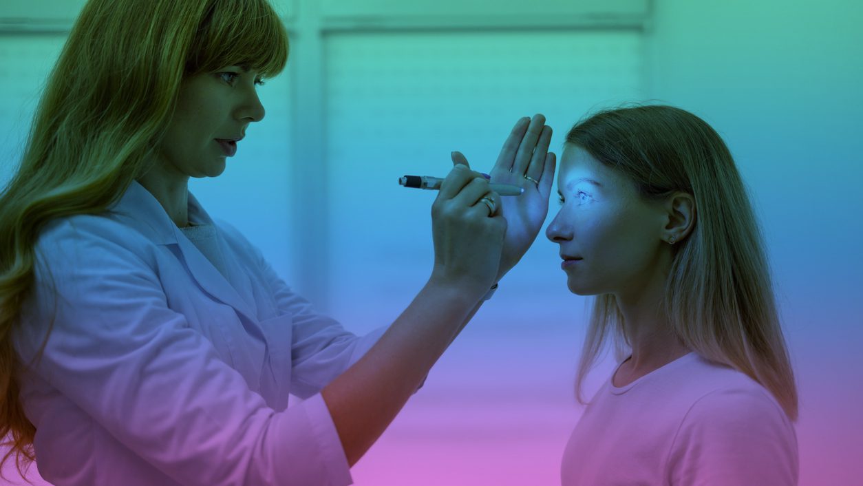
Pupillary Light Reflex
The pupillary light reflex is a neurological process that occurs when the eyes are stimulated by changes in light intensity. Through this reflex, the pupils constrict when exposed to bright light and dilate when exposed to darkness, in order to regulate the amount of light entering the eye.
Pupil dynamics are mediated by the autonomic nervous system (ANS), which often becomes dysregulated following concussion [2]. Patients with altered pupil dynamics may have larger pupils, pupils that are very quick to dilate when exposed to darkness, or pupils that are slower to constrict when exposed to light [3]. Their pupils may also be unable to remain constricted when exposed to light for several seconds. These findings typically indicate increased sympathetic nervous system activity, and depressed parasympathetic nervous system activity [3].
The autonomic nervous system also plays a critical role in regulating heart rate variability, and patients with altered pupil dynamics will often also present with other signs of ANS dysfunction such as exertional intolerance, light headedness with positional changes like when moving from sitting to standing, or have their symptoms worsen with physical activity [2]. Addressing ANS dysregulation and reducing excess sympathetic nervous system tone should help to normalize pupil function.
Accommodation
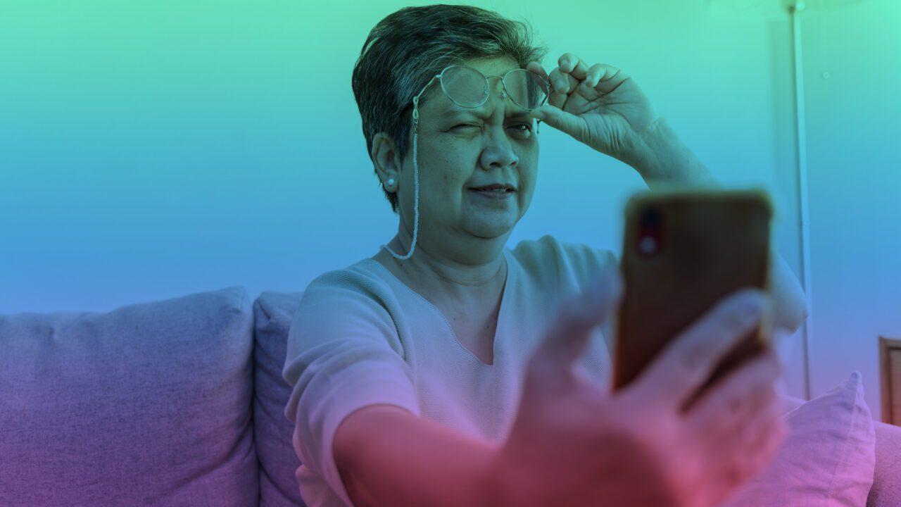
Visual accommodation refers to the ability of the eye to adjust its focus from distant objects to closer ones. Accommodation relies on several parts of the visual system, including neural pathways from the brain and the ciliary muscles in the eye, which work to control the shape of the lens to create a clear image.
Accommodation is measured from the patient’s forehead to the nearest point at which they can maintain focus on a 14 point font test card, with normal accommodation being within 10 cm for a person aged 30 or younger, after which there is an age-associated decline in visual accommodation. When these pathways are not functioning properly, testing accommodation can help identify any deficits that may be present.
Accommodative dysfunction is one of the most common visual changes following concussion injury, with one study of 100 adolescents demonstrating that 51% had accommodative dysfunction following concussion [4].
A patient with accommodative dysfunction may have difficulty focusing on text while reading, or adjusting their vision from a near to far focal point (think of the student looking up at the blackboard or overhead screen, then back down at their paper while taking notes). Patients may also report headache, nausea, blurry vision, dizziness, poor concentration, eye strain or other symptoms while performing these types of tasks.
Accommodative dysfunction can be treated with rehabilitative exercises to work on retraining the associated neural pathways involved in helping the eye adjust its focus.
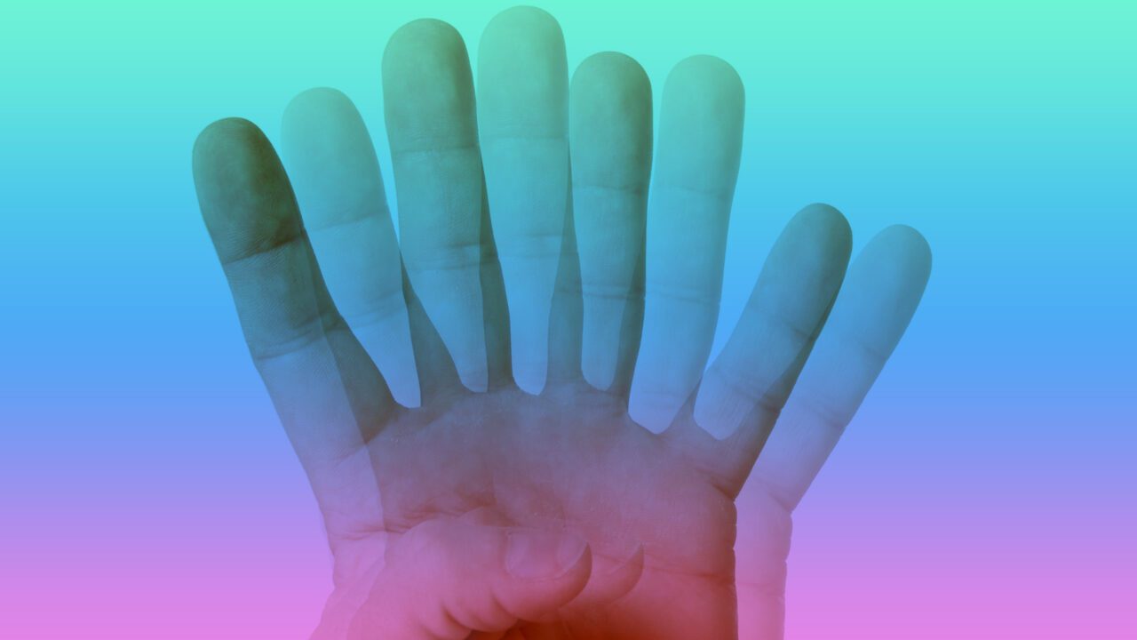
Convergence is the neurological process by which both eyes work together to focus on objects at a close distance (this is also referred to as our binocular vision). Testing of near point convergence is used to assess how well your patient can keep both eyes in alignment when focusing on a near object, and can be helpful in investigating symptoms of double vision following a concussion.
When the eyes are unable to work in tandem to focus on objects at a close distance, this is called convergence insufficiency. According to some recent literature, some amount of convergence insufficiency may occur in as many as 55% of patients following concussion [4]. Convergence is measured as the closest distance from the patient’s nose at which they can focus on a target before the target doubles, with normal convergence being approximately 5 cm or less.
Patients with convergence insufficiency will often report symptoms such as double vision, eye strain or fatigue, headaches, difficulty reading and concentrating, and may also have trouble with depth perception. Additionally, patients with convergence insufficiency or other oculomotor dysfunction may report memory problems associated with reading. While this may appear on the surface to be a cognitive or mental processing issue, it is often a product of reduced visual processing due to the inability of the eyes to work efficiently together.
In many cases, convergence insufficiency can be treated with rehabilitative exercises, but in some instances may require additional assessment and intervention by another healthcare provider such as a neuro-optometrist.
For more information on testing for convergence insufficiency, see our post on the VOMS assessment.
Smooth Pursuits
Smooth pursuits, or eye tracking, refers to the process of being able to follow an object with the eyes as it moves through the visual field. Smooth pursuits rely on neural pathways from the brain and muscles in the eye that work together to help maintain focus on a moving target. Oculomotor dysfunction following a concussion injury can impair a patient’s smooth pursuits.
A concussed person with smooth pursuit deficits may have difficulty tracking a moving object, such as someone running across the room. They also may have issues keeping up with movement in sports or activities that require fast visual tracking. In addition to difficulty with these types of activities, patients may experience symptoms of headache, dizziness or nausea while attempting to perform them. Smooth pursuits impairments can often be addressed with rehabilitative exercises to work on retraining the neural pathways between the brain and the eye muscles that control their movements.
Saccades refer to the fast, precise eye movements that enable us to quickly shift our focus from one point in the visual field to another. Saccadic dysfunction is common following concussion injury, and often leads to difficulty with reading or tracking moving objects.
Saccadic testing is used to assess how well a patient can make large and small jumps between different points in the visual field. Patients with saccadic deficits may experience difficulty scanning lines of text when reading or may frequently lose their place on the page, and have trouble tracking moving objects. In addition to these issues, patients may also report headache, fogginess, dizziness or nausea while attempting to perform tasks that rely on saccades.
Saccadic deficits can often be treated with rehabilitative exercises to help retrain the neural pathways associated with saccadic movement and accuracy.
For more information on testing for saccadic deficits, see our post on the VOMS assessment.
Gaze Stability
Gaze stability is the ability of our eyes to remain stable while focusing on a single point in the visual field. It relies on multiple neural pathways from the brain to help keep our vision steady, so that any movement or disturbances doesn’t cause us to lose our focus. The vestibular ocular reflex plays an important role in stabilizing gaze during head movements by producing an equal and opposite movement of the eyes.
Gaze stability testing can help identify deficits in this ability following a concussion, which can manifest in the form of nystagmus (involuntary eye movements) or problems with visual tracking. Patients may also experience dizziness, headache, nausea, or fatigue when attempting to perform tasks that require gaze stability. Treatment for gaze instability typically includes rehabilitative exercises to help retrain the vestibular ocular reflex and improve visual-vestibular integration.
For more information on testing for gaze instability, see our post on the VOMS assessment.
Visual Motion Sensitivity
Visual motion sensitivity (VMS) is the ability of our visual and vestibular systems to process and respond to changes in movement. This includes tracking fast-moving objects, perceiving depth while moving, or recognizing patterns between different motions. Processing of visual motion integrates input from the visual system, vestibular system, and proprioceptive systems. As such, healthcare providers should assess concussed patients with visual motion sensitivity for vestibular or cervical spine issues, as these may also be factors.
VMS deficits can affect a patient’s ability to perform activities such as playing sports, driving a car, or playing video games. Patients with VMS impairments may experience dizziness, headaches, nausea, eye strain, difficulty concentrating while performing tasks that involve motion, and often report that their concussion symptoms get worse when in busy environments such as sporting events, shopping centres and grocery stores.
VMS deficits can often be addressed with rehabilitative exercises in a physical therapy or rehab setting, and with the use of habituation and desensitization activities in a patient’s day-to-day endeavours.
For more information on testing for VMS, see our post on the VOMS assessment.
Visual Midline Shift
Visual midline shift occurs when there is a mismatch between visual and spatial information processed by the brain, specifically as it pertains to the proprioceptive base of support. It can occur following concussion injuries, more serious traumatic brain injuries, and other insults to the brain such as a stroke.
Patients with a visual midline shift experience an alteration in their sense of position in space, with their perception of the midpoint in the visual field having shifted left, right, anteriorly or posteriorly. Some individuals may present with significant postural deviations that have occurred to account for the change in perception of the visual midline. Those with a midline shift to the right, for example, may present with a right-sided lean, and have associated balance problems or alterations in gait, such as drifting to the right while walking. Patients may also be left feeling as though the floor is slanted, and will compensate accordingly with postural changes.
It is important to note that postural deviations may also be the result of proprioceptive dysfunction in the cervical spine, and an assessment of the neck should always be performed to rule this in or out. More obvious cases of visual midline shift may be assessed in a clinical setting by a rehab professional, but more subtle cases may require neuro-optometric evaluation to arrive at the diagnosis. Cases of visual midline shift may also require neuro-optometric intervention such as yoked prisms in order to correct the dysfunction.
Central-Peripheral Integration
Central-peripheral integration refers to the ability of our visual system to process and respond to stimuli from both the center and outer portions of the visual field. Our central vision is used consciously and is responsible for what we are intentionally looking at. It includes our visual acuity and seeing fine details, and is processed by the brain more slowly. Our peripheral vision processes information about our surroundings, our location in space, where objects are located relative to ourselves, how objects may be moving and at what speed, as well as how we would like to move through space.
Processing by our peripheral vision happens unconsciously, and at a rate much faster than our central vision. It allows us to anticipate things before they happen, and works very closely with vestibular and proprioceptive systems. Our central and peripheral systems must be able to work together seamlessly for many day-to-day activities, such as driving (we have to be able to pay attention to the road in front of us, while being aware of other vehicles on the road such as those in the lane next to us).
Following concussion injuries, our visual system may have difficulty with integrating input from our central and peripheral systems. Often times, our peripheral awareness is reduced and our central vision becomes overly tunneled. Patients experiencing this phenomenon will have normal visual acuity, but may struggle to orient themselves in space or have difficulty processing things coming at them from the sides. It’s important to note that these patients do not have a true loss of their peripheral visual field, but rather reduced processing of what their peripheral vision is taking in.
While there are no specific tests in a rehabilitation setting to diagnose reduced peripheral processing, a detailed patient history can help give you clues about how well the central and peripheral visual systems are working together. Assessing visual fields to confrontation can be performed in your clinical practice to rule out visual field deficits that may indicate a more serious brain injury.
The Impact of the Cervical Spine on Visual Function
The cervical spine and the visual system are intricately connected and are both a part of the sensorimotor feedback loop. The sensorimotor feedback loop is a complex but essential network that helps us to interact with and perceive our environment, and includes input from the visual system, vestibular system, and somatosensory input from different regions of the body, including the cervical spine. This connection is essential for everyday activities such as reading, sports, and driving.
The cervico-ocular reflex induces eye movement in response to proprioceptive feedback from the neck and as such injuries to structures in the neck can cause dysfunction in the visual system [5]. When performing vision tests on your concussion patients, it is important to assess for contributions from the cervical spine. This can be done easily by evaluating the patient’s visual performance while in different head and neck positions.
Common examples of this include testing smooth pursuits, saccades, accommodation and convergence with the head rotated to each side. If the patient’s performance on any of these tests is poorer with the head and neck in a rotated position when compared to the neutral position, this indicates a cervical spine contribution to their visual dysfunction [5]. In such cases, appropriate treatment of the underlying neck pain and cervical spine dysfunction will be necessary in order to help resolve the patient’s vision symptoms.
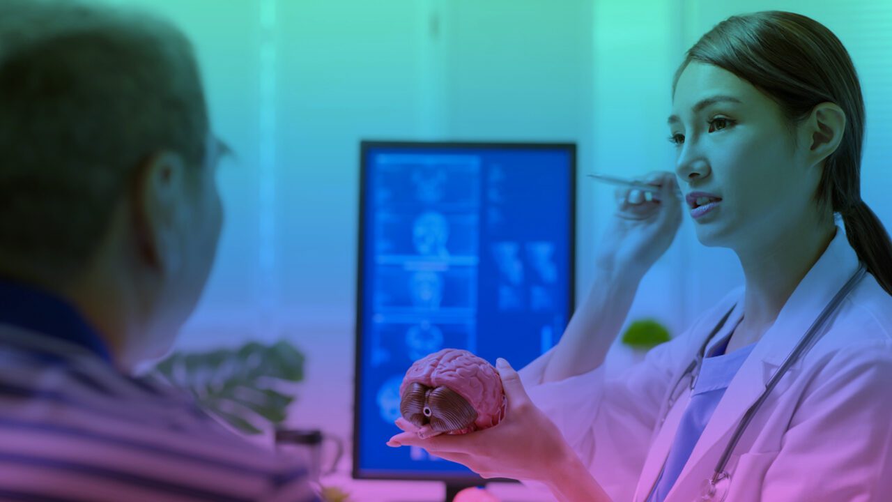
The Role of Neuro-Optometry
Neuro-optometry is a specialty of optometry that focuses on the assessment and treatment of vision-related problems caused by neurological deficits. It combines elements of both traditional optometric care with rehabilitative techniques to help patients recover and/or optimize their vision following such events as traumatic brain injury, stroke, and concussion.
Neuro-optometric assessment may be warranted in your concussion patient depending on the extent of their visual dysfunction and associated symptoms, as well as their response to visual rehabilitation that you provide. For example, patients with significant accommodative dysfunction or binocular vision issues that have not responded to your rehabilitative exercises may benefit from a neuro-optometric referral for further assessment.
Using special in-office equipment and other assessment tools, a neuro-optometrist will perform additional diagnostic testing to assess for vision deficits and concussion related vision problems in order to determine the cause of ongoing visual symptoms.
In some cases, lenses containing different types of filters or prisms may be necessary to help address a patient’s vision problems and symptoms. Neuro-optometrists may also recommend a formalized vision therapy treatment program that includes various in-office and at-home vision rehabilitation exercises to help the brain use the eyes more efficiently during visual tasks. Interventions such as light therapy, which can also help to address sleep problems, may also be utilized [6]. Common post-concussion vision conditions where neuro-optometric intervention may be particularly helpful include visual midline shift syndrome, poor central-peripheral integration, visual snow syndrome, and persistent photosensitivity, among others.
Wrapping It Up.
Visual disturbances can be a common symptom of concussion and other traumatic brain injuries and is one of the most common contributing factors to post concussion syndrome. As a rehab professional working with concussion patients, it is important to assess the patient’s visual system thoroughly in order to identify any potential contributing factors that may be worsening their symptoms. Proper evaluation of both central and peripheral aspects of vision as well as vestibular and cervical spine contributions are essential for developing a comprehensive approach to managing concussion-related visual complaints. In some cases, referral to a neuro-optometrist may be warranted in order to obtain specialized assessments and treatments for helping patients recover from their vision deficits.
By being aware of the potential consequences of head trauma on visual function, rehab professionals can help to provide more comprehensive, individualized care to their concussion patients, leading to improved outcomes for those patients suffering from persistent visual disturbances after their injury.
References
Gunasekaran, P., Hodge, C.,Rose, K., & Fraser, C. (2019, August 1). Persistent visual disturbances after concussion. Australian Journal of General Practice, 48(8), 531–536. https://doi.org/10.31128/ajgp-03-19-4876
Okutucu, S., Civelekler, M., Aparci, M., Sayin, B. Y., Tanalp, A. C., & Oto, A. (2015, March). OP-166 Assesment of the Relationship Between Dynamic Pupillometry and Exercise Heart Rate Recovery among Healthy Subjects. The American Journal of Cardiology, 115, S73. https://doi.org/10.1016/j.amjcard.2015.01.317
Truong, J. Q., & Ciuffreda, K. J. (2016, August 19). Comparison of pupillary dynamics to light in the mild traumatic brain injury (mTBI) and normal populations. Brain Injury, 30(11), 1378–1389. https://doi.org/10.1080/02699052.2016.1195922
Fraser, C. L., & Mobbs, R. (2021, August 30). Visual effects of concussion: A review. Clinical & Experimental Ophthalmology, 50(1), 104–109. https://doi.org/10.1111/ceo.13987
Cheever, K., Kawata, K., Tierney, R., & Galgon, A. (2016, December 1). Cervical Injury Assessments for Concussion Evaluation: A Review. Journal of Athletic Training, 51(12), 1037–1044. https://doi.org/10.4085/1062-6050-51.12.15
Killgore, W. D., Vanuk, J. R., Shane, B. R., Weber, M., & Bajaj, S. (2020, February). A randomized, double-blind, placebo-controlled trial of blue wavelength light exposure on sleep and recovery of brain structure, function, and cognition following mild traumatic brain injury. Neurobiology of Disease, 134, 104679. https://doi.org/10.1016/j.nbd.2019.104679
Dr. Bradford is a chiropractor and a graduate of the Canadian Memorial Chiropractic College in 2017. He began pursuing additional training in concussion rehabilitation after working with competitive cheerleaders and observing significant gaps in the knowledge delivered in the chiropractic curriculum.
Dr. Bradford now works as part of a multidisciplinary team at Altum Health, with a focus in concussion rehabilitation following workplace injuries and motor vehicle accidents. He also operates a small practice out of his home in Cambridge, Ontario where he grew up.
Dr. Bradford’s approach to patient care includes education, myofascial release, acupuncture, joint manipulation and physical rehabilitation, empowering patients to play an active and informed role in their recovery.