Concussions can happen to athletes of any age participating in all kinds of sports. From hard hits in hockey and football to accidents in gymnastics and soccer, concussions are an…
Greater Toronto Area’s Leading Multidisciplinary Clinics for Concussion Recovery
Concussions are a common form of traumatic brain injury, often caused by blows to the head. They can result in an array of physical, cognitive, and emotional symptoms that may…
Why You Should Never Skip the Neck in Concussion Evaluation & Rehab.
Every patient with a concussion is a patient with a neck injury. Let me explain. A concussion results when the brain undergoes such rapid acceleration-deceleration that shearing and stretching of…
Rethinking Concussion Recovery: The Case for Active Rehabilitation Concussions are more than just a bump on the head, or “getting your bell rung”; they are a type of mild traumatic…
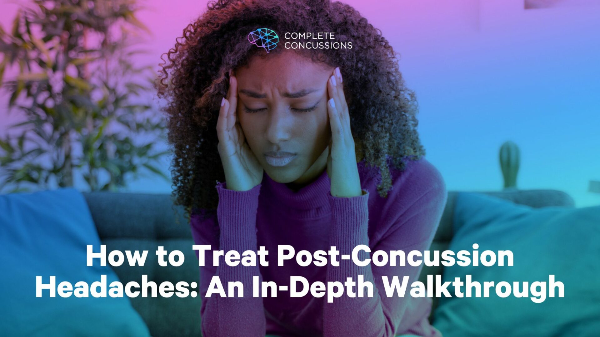
Though common, headaches can be complex and elusive manifestations that can be difficult to treat. In general, there are over 150 kinds of headaches, from the dreaded “brain freeze” (yes, theNational Institutes of Health includes this among its headache types) to the even more dreaded migraine. This vast diversity of headache types implies a wide variety of possible causes.
For clinicians who assist headache sufferers, this can make treatment a daunting task because knowing how to effectively treat something depends very much on knowing, first, what you are treating. This is true all the more for those who manage concussions, since concussion itself is a complex entity and the causes of headache in concussion sufferers can be explained by multiple phenomena.
This article serves as a guide for clinicians who wish to provide the best evidence-based care for patients with post-concussion headaches.
Headaches are one of the most typical symptoms reported after concussion. That being said, there is no such thing as a “concussion headache.” Rather, there are headaches that result from a concussion. Thus, it is better to call them concussion-related headaches.
Concussion-related headache disorders may be caused by dysfunction in a number of physiologic domains:
the nervous system
the visual system
the circulatory system
the gastrointestinal system
the endocrine system
the musculoskeletal system
For some patients, dysfunction in only one of these systems may be the culprit. For many, headaches will result from dysfunction in multiple regions. For example, a patient may be experiencing neck dysfunction secondary to a whiplash injury and blood flow abnormalities to their brain due to autonomic dysregulation. Both of these irregularities may result, or contribute to, headaches.
So, how do we offer treatment for something so potentially complex and multifaceted?
For clinicians, the precise cause of a patient’s post-concussion headaches—as common and predictable as the symptom might be—can be difficult to discern. But while the exact cause is not always clear at the onset, the most probable causes are. For this reason, the best way to treat a patient’s post-concussion headaches is to take a broad, careful, comprehensive, systematic, multimodal approach.
A Broad, Careful, Comprehensive, Systematic, Multimodal Approach.
So, what does this “broad, careful, comprehensive, systematic, multimodal approach” to treatment of concussion-related headaches look like in practice?
First—keep “first things” first. Since treatment is inextricably linked with the results of history-taking and physical examination, the three primary steps that precede and ultimately determine how best to treat concussion-related headaches include:
Step 1: Take a thorough history.
Step 2: Conduct a focused physical exam.
Step 3: Decide top differential diagnoses.
From here, we move to our fourth step which grows organically and climactically out of the prior steps: treat accordingly. So, our broad framework for treating headaches includes:
Step 1: Take a thorough history.
Step 2: Conduct a focused physical exam.
Step 3: Decide top differential diagnoses.
Step 4: Treat accordingly.
A good treatment plan is not something formulated arbitrarily, nor is it based solely on a practitioner’s preferences. A treatment plan should be a logical conclusion from more preliminary, carefully executed steps. So, steps 1-4 must come as a package deal.
For patients with post-concussion headaches, there is a critical distinction you will need to make. While all concussion-related headaches are post-concussion headaches, not all post-concussion headaches are concussion-related. Not every headache experienced after a concussion may be directly related to the head injury. So,
a) Determine the nature of your patient’s post-concussion headaches.
b) Determine your patient’s pre-concussion headache history.
In fact, time could be the only thing relating a post-concussion headache to a concussion. Take Tom (an imaginary patient), for example:
Tom suffered a concussion yesterday and is suffering from a tension-type headache today. To be sure, there is close temporal proximity between his head injury and his headache. At a glance, it seems obvious that this headache is related to the blow to the head he sustained yesterday. But what if Tom suffered from chronic tension-type headaches before sustaining his head injury? If so, it may be that some—or all—of his post-concussion headaches have no direct link to the brain injury he just sustained.
It is the clinician’s task to determine whether a patient’s post-concussion headaches are concussion-related. This is incredibly important, especially when making decisions concerning their return to school, work, and/or sport.
This distinction between post-concussion headaches and concussion-related headaches also highlights the importance ofbaseline testing. If a clinician has a valid baseline test at his disposal for a given patient who has suffered a brain injury, he will much more easily be able to determine the clinical significance of his patient’s post-injury symptoms, including headaches.
And of course, taking a good history will include ascertaining frequency, duration, intensity, character, relieving and aggravating factors, associated symptoms, and so on. The success of the steps that follow depend on this first step being done with thoroughness and precision.
Take your time.
Ask all the questions.
Does the character, intensity, frequency and duration of your patient’s post-concussion headaches, and her associated symptoms, match those of her pre-concussion headaches? Let’s say they don’t. Her headaches are guilty until proven innocent—they are concussion-related.
Step 2: After a Thorough History, Conduct a Focused Physical Examination.
A thorough, focused physical exam should include the following steps:
Begin with observation.
While we don’t want to mistake horses for zebras, we should always rule out worst case scenarios first. Especially if this is your patient’s first visit to a care provider shortly after sustaining a concussion, look for visible red flags like “racoon eyes,” battle sign, evidence of blood or clear fluid at nose/ears, or visible deformation of face/head. Assesslevel of consciousness. Further, look for bruising, lacerations, or abrasions visible in the head or neck areas.
You will also want to note more typical observations like poor posture or postural asymmetries that may contribute to headaches associated with neck or back dysfunction.
Conduct a good neurological exam.
This should include relevant cranial nerve, ophthalmic, and cerebellar tests.
For an example of a comprehensive cranial nerve exam, clickhere. While the cranial nerve exam will assess some aspects of the visual system, a further ophthalmic exam should be undertaken to assess smooth pursuit, saccades, VOR, convergence, and accommodation as these are commonly affected by concussion. Further,cerebellar tests are easy to do and can quickly indicate whether there has been structural damage to the brain.
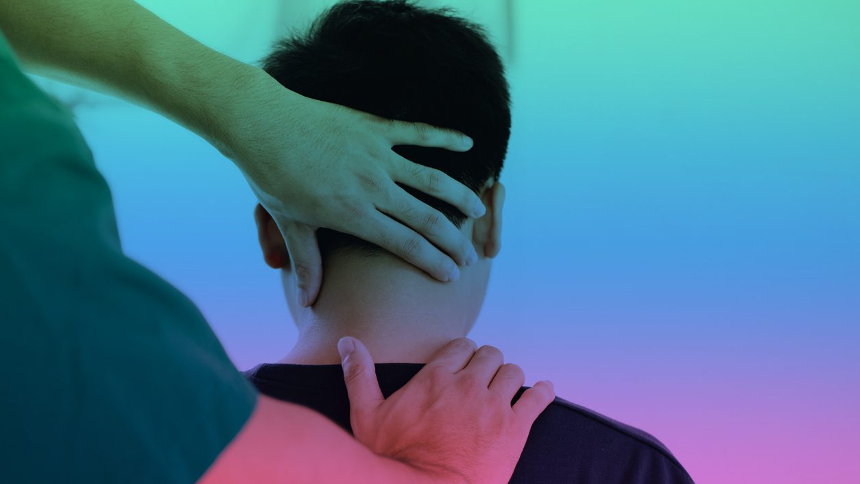
Conduct an orthopaedic assessment of neck and upper back.
First, palpate for midline tenderness at the cervical spinous processes and ensure the patient is able to actively rotate their cervical spine at least 45 degrees bilaterally. Following an event causing head injury, if such c-spine tenderness is present or the patient is unable to rotate their neck bilaterally to 45 degrees, this merits the ordering of x-rays to rule out more sinister pathology (according to theCanadian C-Spine Rule).
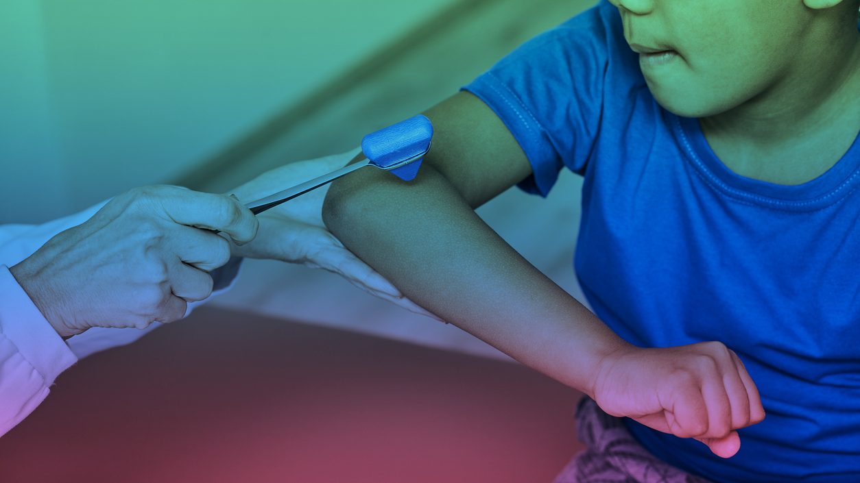
Refer for imaging, if warranted.
Your patient’s signs and symptoms revealed through your history and examination may indicate the need for further diagnostic tests, including radiographs and/or advanced imaging. All clinicians treating patients with concussion should be familiar with guidelines like theCanadian C-Spine Rule andCanadian CT Head Rule.
Neuroimaging is not indicated in patients with recurrent headache with the clinical features of migraine, normal neurologic examination findings, and no red flags.
Step 3: Decide Top Differential Diagnoses
Now, there are two categories of headaches: primary and secondary.
Primary headaches are idiopathic, and typically occur for no specific reason nor do they occur as the result of some other underlying cause. Examples include migraine headaches and tension-type headaches.
Secondary headaches occur directly as the result of underlying causes (usually of the head or neck). Most headaches are not secondary. An example of a secondary headache is a headache caused by an intracranial hemorrhage.
Your first priority as a healthcare provider managing patients with traumatic brain injuries is to rule out anything sinister. Thus, if your history and examination point toward a potentially life-threatening diagnosis, you must act quickly and get into emergent care immediately.
Is your patient’s headache a secondary headache?
Though rare,secondary headaches can be a warning sign of a more serious underlying condition. The SNOOP mnemonic may be used to screen for serious underlying causes of headache. SNOOP stands for:
S – systemic signs and symptoms
N – neurologic symptoms or signs
O – onset sudden
O – onset after the age of 40 years
P – change of headache pattern
These details should be ascertained during your history and physical exam. Since 2003, when the SNOOP mnemonic was first proposed for primary care practitioners, more signs and symptoms have been added to the list. You can access the full listhere.
Once you have ruled out secondary causes of your patient’s headaches, you will determine which primary headache is best indicated by the results of your history and physical exam.
Here are the diagnostic criteria for some of the more common primary headaches.
Migraine with/without aura
The famed neurologist and science writer Oliver Sacks writes inMigraine, “The cardinal symptoms of common migraine are headache and nausea . . . [but] great variability of symptoms is characteristic, not only of attacks in different patients, but between successive attacks in the same patient.” Migraine is a book-length reflection upon the many different—often perplexing—manifestations of this type of headache he had encountered in his practice.
The common migraine is most often unilateral, temporal (“behind the eye”), and usually lasts between 4-72 hours. They are moderate to severe intensity, often debilitating, and can have a pulsatile quality. Nausea or vomiting is often, but not always, present. The patient may report sensitivity to light (photophobia) and/or sound (phonophobia). Often, only resting in a quiet, dark room will relieve symptoms.
Migraine headaches may also be associated with an “aura.”The International Classification of Headache Disorders describes an aura as consisting of “visual and/or sensory and/or speech/language symptoms, but no motor weakness, and is characterized by gradual development, duration of each symptom no longer than one hour, a mix of positive and negative features and complete reversibility.”
Often migraine sufferers will describe which auras are typical for them. Some may experience an aura only some of the time.
Some who suffer from migraine headaches do not experience an aura at all.
Patients who have been suffering from migraines at a frequency of more than 15 days per month and a duration of over 3 months are classified as “chronic” migraine sufferers. Otherwise, migraine is classified as “episodic.”
Tension Headaches
Unlike migraines, tension-type headaches are typically bilateral. Patients may use words like “pressure” or “band-like” to describe their symptoms. These headaches may last minutes to days, may be associated with pericranial tenderness on palpation, and are mild or moderate in intensity. Episodes are often frequent for patients who suffer from tension-type headaches.
Also, in contrast to migraines, tension headaches do not produce nausea or vomiting, nor is sensitivity to light and/or sound reported. They are not aggravated by ordinary daily activities and do not interfere with routine activities as migraines do.
If a patient has suffered more than 15 tension-type headaches over a one month period for longer than 6 months, the headache is “chronic.” If less in frequency or duration, the tension headache is classified as “episodic.”
Cervicogenic Headaches
Cervicogenic headaches are secondary headaches caused by neck pathology. The underlying neck condition could be a soft tissue or joint disorder.
Like migraines, cervicogenic headaches usually affect one side of the head or face. Frequently, they begin at the suboccipital region of the neck and radiate unilaterally to the eye or the temple. Though the cervicogenic headache may present in similar fashion to a migraine, there are some differences to watch for. Unlike migraine, photophobia and phonophobia are rarely associated with cervicogenic headaches. Further, cervicogenic headaches do not tend to cause nausea or vomiting, nor are they as debilitating as migraines.
The hallmark signs of the cervicogenic headache are 1) associated neck pain or dysfunction, and 2) aggravation of the headache with neck movement. Clinical indications of neck-related headache include mechanical exacerbation of pain, reduced cervical range of motion, focal neck tenderness, and trigger points that refer to the head.
With these details in mind, you can now formulate a list of differential diagnoses pertaining to the headaches being suffered by your patient. It is important to remember that conditions don’t always happen with the clarity and coherence of a textbook description. So, your differential diagnosis list might evolve over time. The important thing is to narrow down your “best explanations” of the headaches to a few possibilities, and treat accordingly while remaining vigilant of any new rising signs or symptoms that may alter your primary diagnosis and thus your treatment plan.
Step 4: Provide treatment in accord with history and examination findings.
It is important to note the presence of any comorbidities that might influence your treatment decisions. These might include a history of conditions like:
insomnia
depression and/or anxiety
hypertension
asthma
history of heart disease or stroke
When formulating a treatment plan, begin by making some basic, preliminary recommendations (diet, hydration, exercise, sleep) as part of your treatment plan for your patient with post-concussion headaches.
As you’ll recall, concussion injuries may cause dysfunction in a number of physiologic domains:
the nervous system
the visual system
the circulatory system
the gastrointestinal system
the endocrine system
the musculoskeletal system
Dysfunction in any of these areas may cause or exacerbate post-concussion headaches. As we have noted, it is often difficult to ascertain—especially in the initial days of treatment—which domains are most affected. Due to the etiological ambiguity that often exists in the beginning stages of concussion care, it is reasonable to initiate some degree of therapeutic intervention in all domains.
Your preliminary treatment recommendations, though broad and basic, will benefit all physiological systems simultaneously. This is the foundational part of any further focused treatment strategy you employ.
Preliminary Treatment Recommendations
Clean up the diet.
Recommend supplements.
Optimize hydration.
Exercise below symptom threshold
Improve sleep quality.
These basic recommendations alone, especially when implemented together simultaneously, will provide a deceivingly powerful aid to recovery. Each recommendation is likely to positively enhance function and repair in all affected systems and tissues.
1. Clean up the diet.
Food is medicine. Simpledietary changes can have a hugely positive impact on concussion symptoms like headaches:
Recommendations include avoiding/reducing foods like:
alcoholic drinks
sweets
preservatives/processed foods
gluten (especially white breads and pastas)
bad oils (vegetable, corn, safflower, margarine, canola, soy, peanut)
dairy products
These foods are pro-inflammatory and can be replaced with healthier options like organic fruits and vegetables, fresh fish (especially salmon, mackerel, herring), and free range, grass fed meats.
Increasing “good” fats may also be beneficial (eg., olive oil, coconut oil, flax seed oil, avocado, almonds, etc) for a recuperating nervous system. These changes may help to offset an ongoing inflammatory response and reduce headache symptoms.
Increasing fermented foods like sauerkraut, kimchi, and kefir can assist in replenishing the gut microbiome with “good” flora, which has tremendous implications for both digestion and brain function. Fermented foods are at least as effective as probiotic supplements—if not better—and they taste far better.
2. Recommend supplements.
Although still in its infancy, there is increasing support for various supplements such as Omega-3 fatty acids, creatine, curcumin, magnesium glycinate, and Coenzyme Q10 (CoQ10). For post-concussion therapeutic protocols, these supplements should be taken at the recommended doses for either 4 weeks for acute cases or 8-12 weeks for chronic cases.
***Keep in mind these are general recommendations and other considerations (age, pregnancy, comorbidities, drug interactions, etc) should be considered before making actual supplement recommendations to patients.
Omega-3 fatty acids are essential for brain development and healthy function. They have also been shown to have an anti-inflammatory effect on the brain. Studies in animal models have suggested that omega-3 supplementation improves cognitive function, reduces nerve swelling, stabilizes cellular energy production and increases nerve repair. Two common forms are EPA and DHA. While both are beneficial, a high DHA supplement is recommended since it isfound more abundantly in the brain. 1.5g of high DHA supplement, twice daily, is recommended.
Creatine monohydrate is another supplement that has shown promise in traumatic brain injury research. Creatine is a molecule stored in the muscles, brain, and other organs that plays a key role in maintaining energy homeostasis. Not only can it replace decreased creatine levels resulting from concussion, but it can reduce symptoms and speed recovery. As onestudy puts it, “There is significant overlap between the neuropathology of mild traumatic brain injury (mTBI) and the cellular role of creatine, as well as evidence of neural creatine alterations after mTBI.” Dosing should be no more than 5g per day.
Curcumin is another supplement that may be beneficial due to its high potential to reduce neuro-inflammation andoxidative stress. To optimize its anti-inflammatory and antioxidant properties, the recommended dose for concussion recovery is 400 mg 3 times daily, with food. To maximize absorption, supplements should be taken with black pepper or healthy fats.
Magnesium plays a wide variety of roles in human physiology, and having adequate levels is critical to an optimally functioning nervous system. Magnesium supplementation has been shown to have atherapeutic effect for headache sufferers, including migraine and tension-type headache sufferers.Studies indicate that magnesium levels plummet with traumatic brain injury, and thus magnesium supplementation may play an important role in post-concussion treatment. Magnesium glycinate is the recommended form as it is easier on the stomach compared to others. Recommended daily dose is 200-400 mg for a post-concussion protocol.
CoQ10 is a molecule produced in the body that aids mitochondria during energy production and is a part of the endogenous antioxidant system. Inmultiple studies, CoQ10 has been shown to have beneficial effects in reducing duration and frequency of migraine attack. Recommended dose is 100 mg, 3 times per day (300 mg total per day).
3. Optimize hydration.
The human body is about 60% water. The brain is over 70% water. It goes without saying, then, just how important hydration is for concussion recovery specifically and human performance in general. Studies indicate that even mild dehydration can impair brain function. For instance, astudy published in The Journal of Nutrition concluded that “in healthy young women, mild levels of dehydration result in adverse changes in key mood states such as vigor and fatigue as well as increased headaches and difficulty concentrating.” Several other qualitystudies have reported similar findings.
In general, it is well established that dehydration can be the sole cause of headaches in otherwise healthy individuals. People recovering from a traumatic brain injury should, all the more, stay properly hydrated.
So, how much water should our patients be drinking? The general recommendation is about 2-3 L per day but challenge your patient to drink at least 3 L per day. You should also encourage your patient to refrain from dehydrators like coffee, tea, energy drinks, and alcohol.
And yes, you might as well acknowledge at the onset that hydrating more—even without the dehydrators—will mean peeing more. Just reassure that this is a small price to pay, annoying as it is, for what is gained in exchange. Dehydration is one of the great enemies of recovery and performance.
Now a pro tip for your patients (and perhaps for you too!):
Remember that an eight hour sleep is an eight hour fast. Unless you’re hooked up to an IV, a good night’s sleep is always going to leave you under-hydrated. Consuming a large shot of water first thing in the morning will get the brain and body on track immediately at the day’s onset, raising the likelihood of healing and performance optimization.
Begin each day, immediately upon waking, by downing a large (.5 to 1 L) drink of water with a pinch of salt. After this, during the first hour of the day—while you pray, or meditate, or read, or plan your day—keep a glass or bottle of water close at hand to sip on.
4. Exercise below symptom threshold.
A common consequence of concussion and associated whiplash injuries to the cervical spine is blood flow dysregulation to the brain. Unimpeded and properly regulated blood flow to the brain (and throughout the body) is known to be an important consideration in headache etiology as well.
Exercise is an excellent way to restore proper blood flow to the brain and, in turn, may be part of (or entirely) the solution to the headaches being suffered by your concussed patients.
Encourage your patient to be as physically active as they can be provided they are not exacerbating their symptoms (though they may still be symptomatic) and as long as they are avoiding any high risk activities that put them at an increased risk for a second impact.
For athletes or people who are ordinarily very physically active, you may choose to run aBuffalo Treadmill Test at 5-7 days post-concussion to determine exactly what your patient’s sub-symptom threshold is (or at what intensity they can exercise before increasing symptoms). This will then inform your exercise recommendations and expedite their recovery and return-to-play.
5. Improve sleep quality.
Not only is lack of sleep associated with headache onset in general, but it is also linked with an increase in headache severity.
The average healthy adult requires 7-8 hours of quality sleep per night. Someone recovering from a brain injury needs more, for it is during sleep—and especially during the deepest phases of a night’s sleep—that most of the body’s restorative processes really take off.
This can be a tricky element of the post-concussion treatment plan because a common symptom of concussion is difficulty sleeping. Given both the importance of sleep for recovery and the common post-concussive predisposition to sleep deprivation, the care provider is obliged to offer advice and reassurance in this critical area.
Here aresome recommendations you may provide to your patients to increase sleep quality:
Get up at the same time every morning.
Avoid caffeine
Exercise in the morning
Lower room temperature during sleep period.
Avoid bright light after supper
Avoid napping during the day (or keep the nap to less than 20 mins)
Take potentially sleep assisting supplements like melatonin or magnesium
With the foundations for recovery in place—diet, hydration, exercise, sleep—you can now provide specialized, evidence-based treatment in accord with whichever headache profile best fits what your patient is experiencing.
As you’ll recall, the systematic process we are following is:
Step 1: Take a thorough history.
Step 2: Conduct a focused physical exam.
Step 3: Decide top differential diagnoses.
Step 4: Treat accordingly.
Primary Headache Treatment Recommendations:
What kind of headache are you treating? Does your patient’s post-concussion headache take on more of a migraine quality? Are they unilateral and associated with nausea and sensitivity to light?
Are they associated with neck pain and made worse by cervical spine movement? Are they bilateral and feel band-like like tension headaches?
Based on whatever you ascertain from the history and physical examination, you will now add a more specific, evidence-based headache treatment protocol to your concussion management plan.
Treatment for post-concussive migraines with/without aura may also include:
Spinal manipulation or mobilization (episodic and chronic)
Massage with focus on upper back, shoulder, neck and head regions (episodic and chronic)
Acupuncture (episodic)
Ice application to neck (episodic)
Keeping a headache diary may help in identifying key triggers (episodic and chronic)
Treatment for post-concussive tension-type headaches may also include:
Cervical mobilization or manual traction (episodic and chronic)
Massage with focus on upper back, shoulder, neck and head regions (episodic and chronic)
Keeping a headache diary may help in identifying key triggers (episodic and chronic)
Ice or heat application to head or neck
Treatment for post-concussive cervicogenic headaches may also include:
Spinal manipulation (one week after whiplash injury) or mobilization
Massage with focus on upper back, shoulder, neck and head regions
Deep neck flexor strengthening exercises
Postural correction/advice
Ice pack application to neck
Final Thoughts.
If a patient comes to you with a head injury and related pain in their head, you have a plethora of treatment options at your fingertips. But remember: your first duty is to check for red flags, that is, to rule out an emergent cause of headache.
Though your patient may be inclined to turn immediately to nonsteroidal anti-inflammatory drugs, reassure them that there are plenty of other conservative options that may help them. (In the beginning hours and days after concussion, pain medications are discouraged anyhow because they can mask concussion symptoms that may be important to be aware of.)
Finally, it is important to remember that treatment is not just something the clinician does. It is a partnership between clinician and patient, and requires an alliance on both ends. You must do your part, but your patients must do theirs as well. Keep an open line of communication with your patients and always be ready to adapt the plan. Further, as you offer your treatment plan and move forward with it, don’t over promise on the anticipated outcomes but assume a balanced posture that exudes both reassurance and realism, optimism and patience.
Almost always, if you remain faithful to evidence-based protocols such as those adopted by the professionals inComplete Concussions clinics, your patients will get better—the headaches go away. People will get their lives back. And when they do, the people you have helped will be more thankful for your assistance than you will ever know.
References
International Headache Society. The International Classification of Headache Disorders: 2nd edition. Cephalalgia 2004;24 Suppl 1:9-160.
Patricios JS, Schneider KJ, Dvorak J, et al Consensus statement on concussion in sport: the 6th International Conference on Concussion in Sport–Amsterdam, October 2022 British Journal of Sports Medicine 2023;57:695-711.
Becker WJ, Findlay T, Moga C, Scott NA, Harstall C, Taenzer P. Guideline for primary care management of headache in adults. Can Fam Physician. 2015 Aug;61(8):670-9. PMID: 26273080; PMCID: PMC4541429.
Bryans, R., Decina, P., Descarreaux, M., Duranleau, M., Marcoux, H., & Potter, B. Clinical Practice Guideline for the Management of Headache Disorders in Adults: www. Chiropracticcanada .ca; 2012 [cited 2018-10-22]. Available from: https://www. chiropractic. ca/wp-content/uploads/2014/09. Headache-CPG-final-Jan2012_English. Pdf.
Pearce KL, Sufrinko A, Lau BC, Henry L, Collins MW, Kontos AP. Near Point of Convergence After a Sport-Related Concussion: Measurement Reliability and Relationship to Neurocognitive Impairment and Symptoms. Am J Sports Med. 2015 Dec;43(12):3055-61. doi: 10.1177/0363546515606430. Epub 2015 Oct 9. PMID: 26453625; PMCID: PMC5067104.
Master CL, Bacal D, Grady MF, et al; AAP Section on Ophthalmology; American Academy of Ophthalmology; American Association for Pediatric Ophthalmology and Strabismus; American Association of Certified Orthoptists. Vision and Concussion: Symptoms, Signs, Evaluation, and Treatment. Pediatrics. 2022;150(2):e2021056047.
Stiell IG, Wells GA, Vandemheen KL, Clement CM, Lesiuk H, De Maio VJ, Laupacis A, Schull M, McKnight RD, Verbeek R, Brison R, Cass D, Dreyer J, Eisenhauer MA, Greenberg GH, MacPhail I, Morrison L, Reardon M, Worthington J. The Canadian C-spine rule for radiography in alert and stable trauma patients. JAMA. 286 (15): 1841-8.
Stiell I, Wells G, Vandemheen K et al. The Canadian CT Head Rule for Patients with Minor Head Injury. Lancet. 2001;357(9266):1391-6.
Do TP, Remmers A, Schytz HW, Schankin C, Nelson SE, Obermann M, Hansen JM, Sinclair AJ, Gantenbein AR, Schoonman GG. Red and orange flags for secondary headaches in clinical practice: SNNOOP10 list. Neurology. 2019 Jan 15;92(3):134-144. doi: 10.1212/WNL.0000000000006697. Epub 2018 Dec 26. PMID: 30587518; PMCID: PMC6340385.
Sacks OW. Migraine: Understanding a common disorder, Berkeley, CA: University of California Press, 1970.
Gupta A, Summerville G, Senter C. Treatment of Acute Sports-Related Concussion. Curr Rev Musculoskelet Med. 2019 Jun;12(2):117-123. doi: 10.1007/s12178-019-09545-7. PMID: 30887284; PMCID: PMC6542872.
Gualano B, Rawson ES, Candow DG, Chilibeck PD. Creatine supplementation in the aging population: effects on skeletal muscle, bone and brain. Amino Acids 48(8), 1793–1805 (2016).
Dean PJA, et al. Potential for use of creatine supplementation following mild traumatic brain injury. Concussion 2017 Jun;2(2): DOI:10.2217/cnc-2016-0016 (2017).
Yablon LA, Mauskop A. Magnesium in headache. In: Vink R, Nechifor M, editors. Magnesium in the Central Nervous System [Internet]. Adelaide (AU): University of Adelaide Press; 2011. Available from: https://www.ncbi.nlm.nih.gov/books/NBK507271/
Institute of Medicine (US) Committee on Nutrition, Trauma, and the Brain; Erdman J, Oria M, Pillsbury L, editors. Nutrition and Traumatic Brain Injury: Improving Acute and Subacute Health Outcomes in Military Personnel. Washington (DC): National Academies Press (US); 2011. 12, Magnesium. Available from: https://www.ncbi.nlm.nih.gov/books/NBK209305/
Sazali S, Badrin S, Norhayati MN, Idris NS. Coenzyme Q10 supplementation for prophylaxis in adult patients with migraine-a meta-analysis. BMJ Open. 2021 Jan 5;11(1):e039358. doi: 10.1136/bmjopen-2020-039358. PMID: 33402403; PMCID: PMC7786797.
Lawrence E. Armstrong and others, Mild Dehydration Affects Mood in Healthy Young Women, The Journal of Nutrition, Volume 142, Issue 2, February 2012, Pages 382–388, https://doi.org/10.3945/jn.111.142000
Haider MN, Leddy JJ, Wilber CG, Viera KB, Bezherano I, Wilkins KJ, Miecznikowski JC, Willer BS. The Predictive Capacity of the Buffalo Concussion Treadmill Test After Sport-Related Concussion in Adolescents. Front Neurol. 2019 Apr 24;10:395. doi: 10.3389/fneur.2019.00395. PMID: 31105634; PMCID: PMC6492460.
Dr. Matthew Nelson is a graduate of the University of Regina (Physical Education, 2007) and the Canadian Memorial Chiropractic College (2014) where he graduated with Clinic Honours and was the recipient of the Istrati Family Memorial Award. He is currently pursuing an MA in Philosophy at Holy Apostles College and Seminary.
Matt played football for the University of Regina Rams for four seasons. Following his time with the Rams, he trained for one year in Bobsleigh Canada Skeleton’s development program. He is owner and clinician at Core Health + Performance in Shaunavon, Saskatchewan. He is married and the father of four children.